CML is often identified through routine blood tests for other medical reasons.
Complete medical history and physical examination are the first investigations.
Hyperleukocytosis with granulocytes in peripheral blood and bone marrow supports CML diagnosis.
BCR-ABL rearrangement establishes diagnosis.
Peripheral Blood Film:
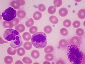
The peripheral blood from a patient with CML shows:
- Borderline anemia with normocytic and normochromic cells.
- Occasional elliptocytes are present (not shown in the image provided).
- Metamyelocytes are present with myelocytes and promyelocytes present also. Blast cells may also be found.
- The neutrophils have a normal morphology and show prominent granulation.
- The basophils appear slightly hypogranulated and are increased along with eosinophils and monocytes.
- There is an increased number of platelets with occasional platelet clumping and large forms present (not shown in image provided).
Diagnostic Procedures for CML:
Signs of Disease |
|
Physical examination |
|
Complete blood count |
|
Blood tests |
|
Cytogenetic analysis |
|
Bone marrow aspiration and biopsy |
|