CBC and Differential
Most important initial test and usually the first laboratory investigation to return abnormal.
Most common finding will be elevated WBC count (anywhere from >15 x 109/L to 50 x 109/L).
10% of patients have “normal” CBC.
Important to recognize that WBC count can also be:
- Normal (5-15x109/L)
- Low (“leucopenia” <5x10⁹/L)
- Very high (“hyperleukocytosis” >50 x 109/L up to >1000 x 109/L)
Patients are often also severely anemic and thrombocytopenic. Reticulocyte counts are very low reflecting inability of the bone marrow to produce new red cells because of leukemic infiltration throughout.
The differential on the WBC will come back with “other cells” or “blast cells” by the automated counter. These may be all (or most) of the white blood cells circulating or only a small proportion.
Important to remember that it is usually not possible to diagnose the type of acute leukemia from just peripheral blood smear (precursor B ALL, T-ALL, or differentiate from AML).
| Investigation | Abnormality usually seen |
Hemoglobin |
Decreased |
WBC |
Increased or decreased or normal |
Differential WCC |
Often neutropenia & blasts |
Platelets |
Decreased NB:
|
The Peripheral Blood Film:
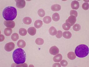
Review of the peripheral blood under the microscope will reveal leukemic “blasts” (the 3 larger purple – blue cells) shown in the figure below.
Normal white blood cells (neutrophils, lymphocytes, monocytes) are absent.
ALL Blasts are characteristically:
- Mainly composed of nucleus and little cytoplasm (high N:C ratio)
- The chromatin in the nucleus is fine, speckled, and immature appearing (like salt and pepper, as opposed to mature chromatin which is denser and more clumping)
- The nuclear contours may be very irregular (folding, convoluted) or more smooth as in this photomicrograph
ALL leukemic blasts:

Other Investigations Important to Order at Time of Initial Diagnosis:
Investigation |
Reason / Abnormality usually seen |
Chest X-ray |
Rule out mediastinal mass from thymic and lymph node involvement (often in T-ALL). |
Group and Screen |
Patient will likely require both blood and platelet transfusions at initial presentation. |
PTT, INR, Fibrinogen |
Usually normal but may be coagulopathic at presentation (more common in AML than ALL) |
Potassium |
Usually normal initially but may be elevated at diagnosis reflecting high cell turnover |
Phosphorous |
Usually normal initially but may be elevated at diagnosis reflecting high cell turnover |
Calcium |
Usually normal but may be low reflecting binding with PO4 if elevated. |
Uric Acid |
Usually high reflecting high cell turnover |
Lactate Dehydrogenase |
Usually high reflecting high cell turnover |
Creatinine |
Often normal but in rare situations may be elevated reflecting either leukemic infiltration of kidney or tumor lysis syndrome occurring before starting chemotherapy (rare). |
Liver Enzymes and Bilirubin |
May be elevated reflecting hepatitis from leukemic infiltration. |
HLA-Typing of siblings and parents |
Ordered by pediatric oncologist in certain situations only where possibility of BMT exists. |
Viral Serologies |
CMV (Cytomegalovirus), VZV (Varicella), HSV (Herpes Simplex Virus), EBV, HIV, Hepatitis B, Hepatitis C, Mumps, Measles, Rubella |
Echocardiogram |
Ordered by pediatric oncologist to evaluate baseline heart function if possibility exists patient will need anthracycline chemotherapy (affects heart, used in high-risk ALL only). |

