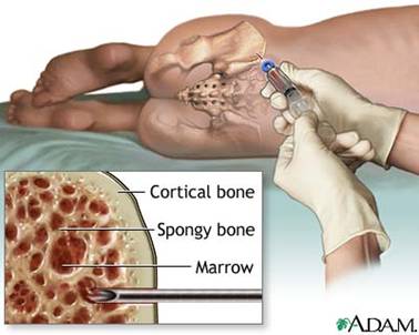
Bone Marrow Aspirate under conscious sedation using an aspirate needle into the posterior superior iliac spine.

Draws the bone marrow out as a liquid that looks like “frothy blood”. Cells are disrupted from their normal architecture.
Available to be sent for:
- Morphological assessment
- Flow cytometry for immunophenotyping
- Karyotype analysis
- Molecular genetic profiling for known genetic changes of ALL
Marrow aspirate most important test as allows for confirmatory diagnosis, characterization of type of acute leukemia (precursor-B, T-ALL, biphenotypic, bilineage, AML, MDS – see below) and offers prognostic information according to genetic abnormalities.
Diagnosis of type of acute leukemia ultimately made by collective review of morphology, immunophenotyping (see below), and genetic information.
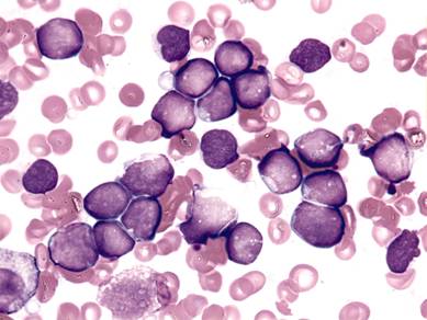
Figure below: Morphologic assessment of newly diagnosed precursor B-ALL from a bone marrow aspirate. Nucleated marrow cells are mostly leukemic blasts of similar appearance.

Bone Marrow Biopsy
Maintains the bone marrow in its native architecture.
Allows estimation of marrow cellularity.
Usually shows clusters of blast cells infiltrating throughout marrow space.
Not universally performed with the aspirate at all centers (center dependent).
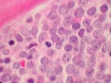
Figure below: Bone marrow biopsy. Note the cells all appear similar (leukemic blasts) and literally pack all of the space in the marrow cavity, making the overall cellularity 100%. Normally marrow should have some “open” space that is made up of fat (giving a cellularity around 70%) and the mix of cells reflecting normal trilineage hematopoiesis.


