Ultrasound scan:
In infants:
- Investigation for infants with an enlarged head circumference and an open anterior fontanelle
- Transcranial ultrasound may identify an abnormal mass
CT Imaging:
Supratentorial PNETs tend to be:
- Sharply demarcated and not infiltrative
- Large, bulky tumors
- Heterogeneous due to the variable presence of hemorrhage, necrosis, calcification, and cysts
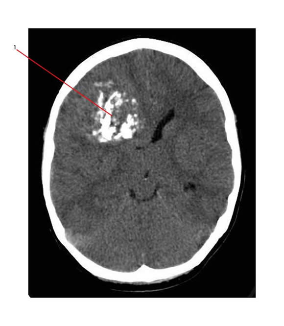
- Intratumoral calcification in 50%
- Solid component appears as a hyperdense mass
- Tumor enhances brightly and heterogeneously with contrast
- Usually associated with minimal or no edema
The CT below shows intra-tumoral calcification (1) in a large supratentorial PNET

MRI Imaging:
This is the "gold standard" to assess the extent of disease (delineates anatomical confines) and the presence of leptomeningeal dissemination.
This investigation helps to distinguish supratentorial PNETs from other tumors.
The tumor is:
- Usually isointense to cortex on FLAIR sequence MR scans
- Solid components have a high signal on diffusion-weighted MR imaging with restricted diffusion on apparent diffusion coefficient map
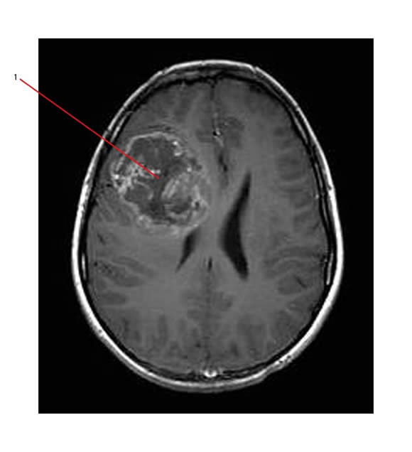
- Heterogeneous mass with relatively well-defined margins, and moderate to intense enhancement with gadolinium.
Different MR sequences:
- T1-weighted MRI
- typically heterogeneously low signal but may be high signal depending on the presence of blood
- T2-weighted MRI
- generally appear isointense to cerebral cortex but may also appear hyperintense
MR Detects intracranial or spinal leptomeningeal dissemination
- 20%–40% of supratentorial PNETs have leptomeningeal spread at presentation
The MR below shows the same large supratentorial PNET as above (1) on MR (T1, post gad sequence):

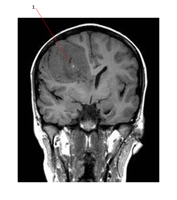
Here is the same tumor (sagittal view)

Link:


