
A complete history and physical examination is always the first investigation.
Imaging studies:
- Ultrasound scan of head (infants with open fontanelle)
- CT head
- MR of head and craniospinal axis
Biopsy:
Surgery would be attempted initially to resect the tumor as completely as possible and biopsy of the primary tumor prior to resection is not generally performed.
Lumbar puncture (LP) to check CSF cytology:
Positive cytology would show "small round blue cells" exactly similar to those found in medulloblastoma (infratentorial PNET).
NB LP would not be done until after tumor resection when the patient's raised intracranial pressure has been treated.
PNET cells in CSF:
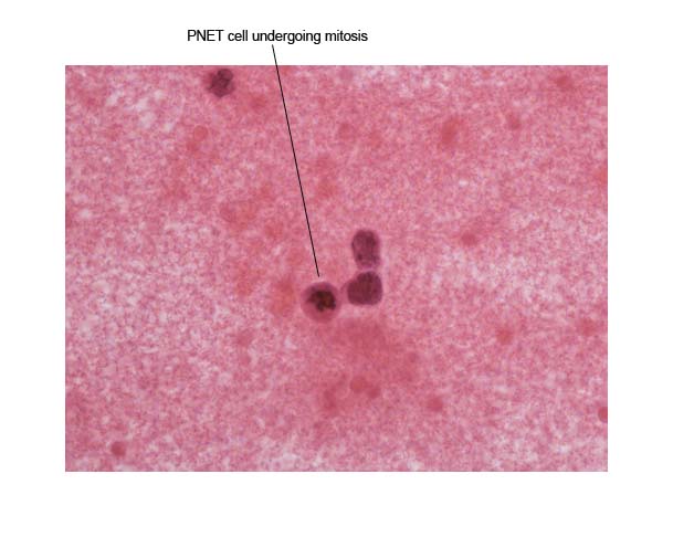
The above slide shows 3 metastatic PNET cells in CSF, one of which is undergoing mitosis.
Other: Metastatic deposits outside the CNS to areas like bone marrow or bone are very rare and unless there are symptoms which are suspicious for metastatic disease, bone marrow biopsy and bone scan would not be routinely performed.