Splenomegaly
The spleen is involved in about 20 - 30% of cases.
Patients are usually asymptomatic.
The frequency of splenic involvement depends on the histologic type of Hodgkin Lymphoma.
Spleen is involved in:
- 15% of lymphocyte predominant
- 35% of nodular sclerosis
- 60% of mixed cellularity
- 80% lymphocyte depleted
The splenic hilar nodes and para-aortic nodes are involved in 50% of Hodgkin lymphoma patients with splenic disease.
In the images below:
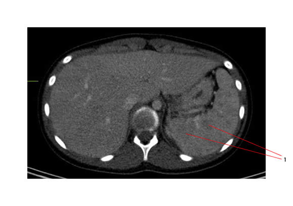
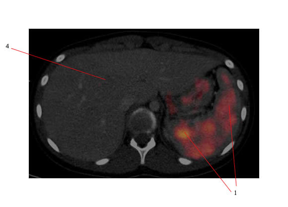
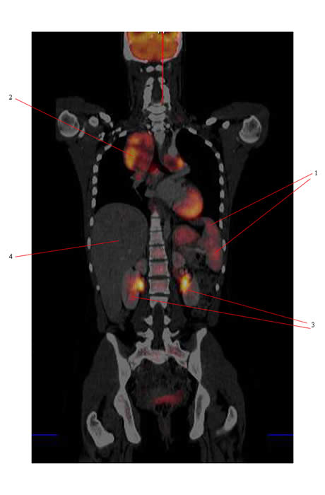
- #1 - nodules of HL involving the spleen
- #2 - particularly large mediastinal HL mass
- #3 - normal kidneys
- #4 - normal liver
Below is the axial CT image showing multiple subtle splenic hypodensities secondary to HL.

Below is the PET/CT (same axial CT image but with the PET data blended in showing that the hypodensities are hypermetabolic).

Below is the coronal blended PET/CT image.

Differential diagnosis of splenomegaly

