The spleen should not be palpable after 3 or 4 years of age.
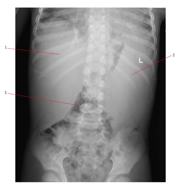
Below is an abdominal film (patient is erect) taken of a child who presented with massive hepatosplenomegaly. #1 points to the liver, #2 points to the spleen and #3 shows bowel loops that have been displaced inferiorly by the organomegaly. This child had T cell acute lymphoblastic leukemia.

Differential Diagnosis of Splenomegaly:
Congenital vs Acquired |
Cysts | Rarely causes mass like abdominal swelling
|
| Hemolytic anemias |
|
|
| Storage diseases |
|
|
| Osteopetrosis | Extramedullary hematopoiesis | |
Inflammatory |
Non-infectious |
|
| Connective Tissue Disease (rheumatoid arthritis) | ||
| Infectious | Viral (EB virus, CMV, HIV, hepatitis A, B & C) | |
| Bacterial (acute and chronic systemic infection, TB, subacute endocarditis, abscess, typhoid fever). | ||
| Rickettsial (Rocky Mountain Spotted fever) | ||
| Protozoal infection (malaria, toxoplasmosis) | ||
| Fungal Infection (systemic candidiasis, histoplasmosis, coccidiomycosis) | ||
| Trauma | Hemorrhage in spleen | Subcapsular hematoma hemangioma, lymphangioma |
Neoplastic |
Malignant | Infiltrative with leukemia, lymphoma (Hodgkin and NHL) |
| Benign |
|
|
| Langerhans cell histiocytosis | ||
| Systemic | Myeloproliferative disorders | Polycythemia vera |
| Congestive splenomegaly |
|
|
| Pre-hepatic or portal vein obstruction |

