MR and CT scan
Generally, low grade astrocytomas cannot be reliably differentiated from more diffuse anaplastic varieties based on imaging alone.
CT scan:
- Enhancement varies from none to extensive depending on the degree of necrosis and cyst formation.
- The borders of well-differentiated tumors often appear isodense to normal brain tissue.
MR scan:
- Astrocytomas are usually:
- Relatively homogeneous mass lesions
- Hypocellular and high in water content
- Low grade astrocytomas may sometimes be differentiated from more malignant astrocytomas on MR as they lack significant peritumoral edema.
- While focal cystic areas may be seen on imaging studies, it may not be possible to distinguish microcystic change from macroscopically cystic regions of tumor in either CT or MRI imaging.
- Contrast enhancement on MRI is not recognized as a reliable indicator of the grade of astrocytoma.
- After contrast administration, well-differentiated astrocytoma has a variable appearance, usually lacking any significant contrast enhancement.
- For juvenile pilocytic astrocytomas, T1 weighted images have a low signal intensity and T2 weighted images have a high signal intensity. There is often an enhancing nodule, or enhancing rim of the cystic component of the tumor.
Examples:
Low Grade
Below is a MR scan (axial T1 image) of a low grade pilocytic astrocytoma (#1).
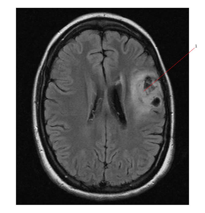
High Grade
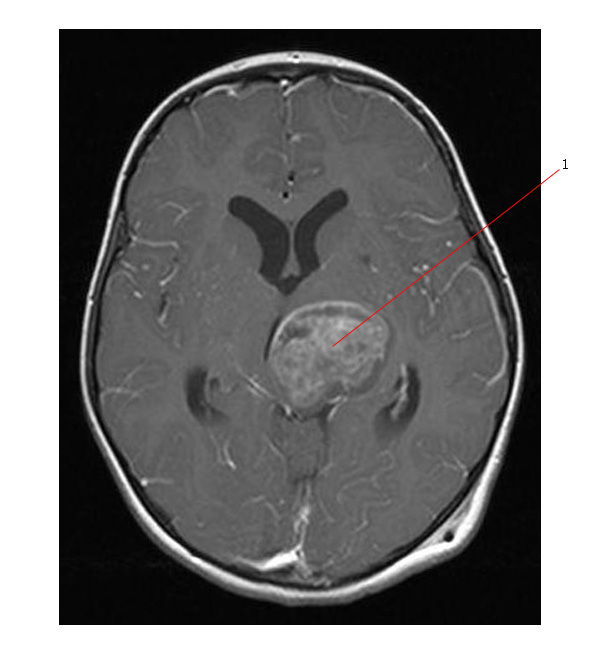
The axial MR image below is of a grade 3 astrocytoma (#1).
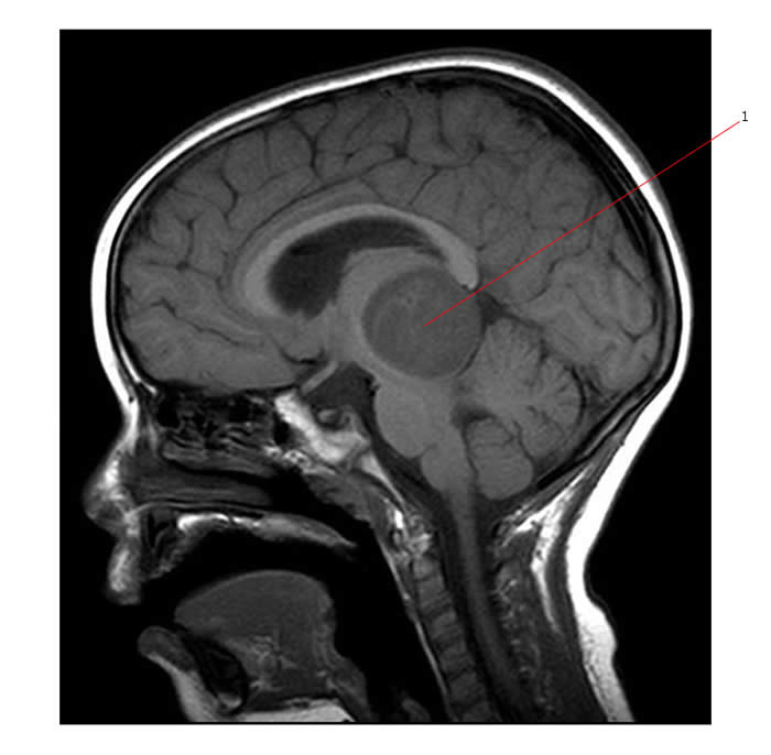
Below is a sagital image of the same tumor (#1).
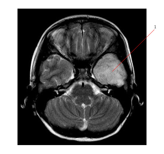
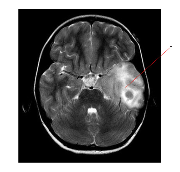
Below is a MR scan (T2, axial) of a glioblastoma multiforme(#1) involving the temporal lobe. This is an aggressive tumor diffusely infiltrating the brain. The child had a very short history of symptoms.
Below is the same tumor at a higher level.
Link:


