Ependymoma
Investigation
An initial thorough history and physical is critical.
Local Disease
The work-up for ependymomas primarily involves CT and MRI.
CT scan is often the initial investigation to assess a child who might have a brain tumor.
MR is used to:
- Assess the extent of local disease in more detail
- Rule out craniospinal metastatic disease
Metastatic Disease
Ependymomas may be associated with leptomeningeal spread.
The risk of craniospinal metastatic disease is 10 - 15% in ependymoma (significantly less than for medulloblastoma).
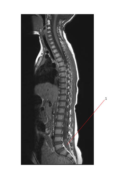
The entire neural axis should be imaged using MRI with gadolinium to look for metastatic disease.
CSF cytology with lumbar puncture should also be performed to determine whether there has been dissemination. This is usually done 21 days after surgery.
Below is a MR scan of the spine showing a metastatic ependymoma deposit (#1) in the lumbosacral region.

Pathology
Surgery is performed and as complete a resection as possible is performed to:
- Confirm the pathological diagnosis
- Achieve local control

