
Ependymoma
Radiology
CT Scan
- Prior to contrast administration, the tumor is predominantly isodense midline posterior fossa mass with variable enhancement.
- Cyst formation is common and readily visible on CT.
- Calcifications appear in 44% of cases, and are either diffuse or coarsely nodular.
MRI scan
- Signal intensity is the same in ependymoma as other intracranial gliomas.
- MR defines the location and gross morphology of the tumor.
- It is important to accurately define the caudal extent of the tumor (for the purposes of resection and subsequent radiotherapy).
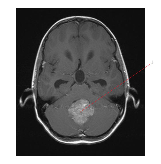
- Ependymomas appear hypointense on T1-weighted images, hyperintense on T2-weighted images, and usually enhance with gadolinium.
- Ependymomas present as a nonspecific heterogenous signal. This heterogeneity in imaging studies can reflect cystic regions, areas of necrosis, hemorrhage, and calcification.
- Gadolinium contrast enhancement occurs in nearly all ependymomas.
The MR (T1 weighted) below shows a midline posterior fossa ependymoma. #1 points to the tumor.
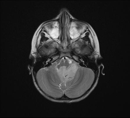
Below is a more extensive tumor arising again in the mid line.
While CT and MRI can give a probable diagnosis, the final diagnosis cannot be made without tissue sampling.
Link: