Ependymoma
Location
Ependymomas arise along the lining of fluid-filled cavities of the brain. In children these tumors occur in the following locations:
- Supratentorial (30%)
- Infratentorial (60%)
- Arise in the ependymal lining of the 4th ventricle
- Spinal (10%)
Infratentorial (Posterior fossa)
Most often midline lesions originating from the ventricular floor.
Posterior fossa is the commonest location and tumors arise from the roof, floor, or lateral resources of the 4th ventricle. Attachment is commonly observed at the level of the obex, where the 4th ventricle ends and the spinal canal begins.
These tumors commonly extend:
- into the foramina of Lushka, growing towards the cerebellopontine angle
- occlude the 4th ventricle and extend cephalad into the aqueduct
- caudally through the foramen magnum and down adjacent to the upper cervical spine
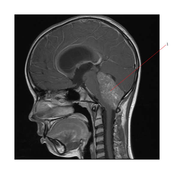
Below is a MR (sag. view) of a posterior fossa ependymoma (#1).

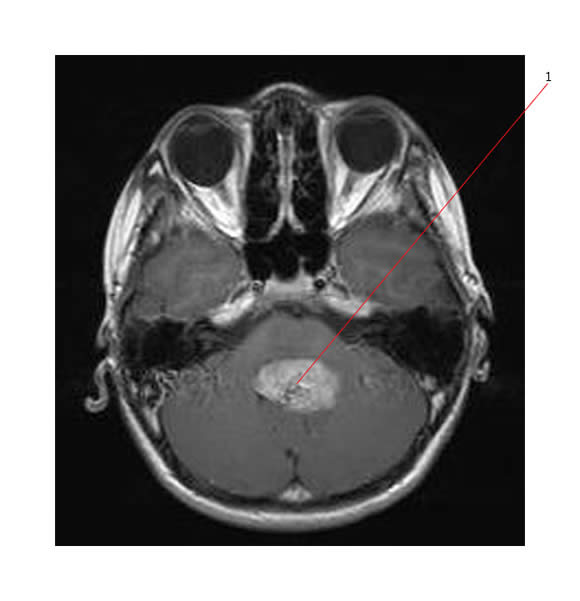
Below is an axial MR (T1 weighted) of a posterior fossa ependymoma (#1).

Supratentorial
Supratentorial ependymomas arise from the ependymal lining of the lateral and 3rd ventricles.
Tumor may be totally or partially intraventricular, and has a predilection for frontal, temporal, and parietal lobes, as well as for the 3rd ventricle.
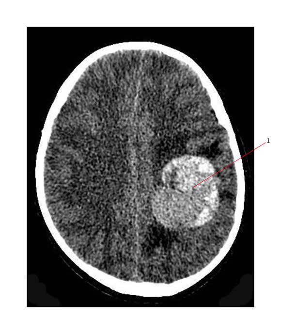
Below is a contrast enhanced CT scan showing a high grade supratentorial ependymoma in the left parietal region (#1). There is significant surrounding brain edema with mild midline shift.

There are also differences between those tumors that arise in adults and those in young children.
- 75% of adult ependymomas arise in the spinal cord.
- In young children, most ependymomas arise in the 4th ventricle.
- With increasing age, supratentorial locations become more common.
Table: Infratentorial vs. Supratentorial Ependymomas
| Infratentorial | Supratentorial | |
| General area | withn the 4th ventricle | Intraventricular or intracerebral
Common locations:
|
| Tissue of Origin | Ependyma (cells lining the ventricles) |
Ependyma |
| Incidence | 2/3 of pediatric ependymomas-more common in children | 1/3 of pediatric ependymomas-more common in adolescents |
| Location of origin | Roof, floor, or lateral recesses of the 4th ventricle | Ependymal lining of the lateral and 3rd ventricles-may be partially or entirely intraventricular |
| Tumor Growth | Common growth patterns:
|
|

