The appearance of the tumor depends on its position and direction of growth.
Disease may be confined to either the chiasm or the hypothalamus.
Smaller nerve and chiasmatic tumors are visually distinct from the hypothalamus, making their site of origin more clear-cut.
These small tumors can be difficult to distinguish from optic neuritis (which can also result in optic nerve enlargement). To distinguish between the two conditions:
- Clinical history is important
- Neuritis is painful
- Follow-up should show resolution of optic nerve swelling in neuritis
CT
This study is rarely used.
CT can be used to identify:
- Total anatomic extent of the tumor.
- Length and width of the optic nerve.
- Papilledema appears on CT as an enlargement of the optic nerve. The plane of the section should be parallel to the optic nerve, as a change in size of the optic nerve or an increased amount of convolutions is not definitively indicative of a tumor.
As these are usually isodense gliomas, intravenous contrast shows little enhancement.
MRI
MR is the gold standard for imaging of these tumors.
Allows for multiplanar visualization of the optic pathway.
- Elongation of the nerve as well as kinking of the nerve may both be indicative of optic pathway gliomas.
- T1 images of optic gliomas appear as isointense, whereas T2 images are of moderate hyperintensity.
- Cysts commonly appear in images of larger hypothalamic lesions.
- Calcification and hemorrhage are rare in these tumors.
- Contrast enhancement appears in half of all cases.
- T2-weighted images of the entire brain are necessary, as this will show the invasion of the tumor along the optic radiations as hyperintensity.
A differential diagnosis of hamatoma is supported by lack of interval growth and possible absence of contrast enhancement.
The presence of tumor extension is supported by the confinement of the tumor to optic pathways, tumor enhancement, and a significant mass effect.
EXAMPLES
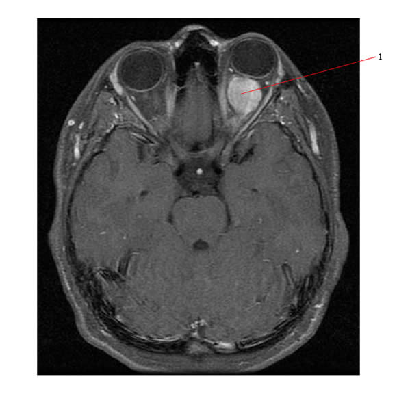
The axial MR below shows a unilateral optic nerve glioma (#1)
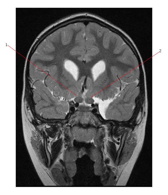
Below is a Coronal T2 image that demonstrates bilateral optic nerve gliomas (#1 and #2 point to these gliomas) in the setting of NF1.
Link:
