Hereditary Factors
Neurofibromatosis
Optic tract gliomas are linked to the hereditary disorder neurofibromatosis type 1 (NF1)
- Commonest type of neurofibromatosis (about 80%)
- Also called von Recklinghausens disease.
- Autosomal dominant hereditary disorder.
- In half of all patients there is a family history and half have a spontaneous mutation.
- Severity of symptoms varies significantly between different individuals (variable expression).
- The NF1 gene has been mapped to chromosome 17q11.2.
- NF1 is associated with the development of:
- optic nerve gliomas
- other CNS tumors
- soft tissue sarcomas (malignant peripheral nerve sheath tumors) later in life
Two of the following must be present to make the diagnosis of NF1.
- Optic nerve glioma
- Axillary freckling (Crowe’s sign)
- Café –au-lait spots (six or more and have to be over 0.5 cm in children and over 1.5 cms in adults)
- Two or more neurofibromas and one plexiform neurofibroma
- Two or more hamartomas of the iris (Lisch nodules)
- First degree relative affected with NF1
- Bony lesion – thinning of long bone cortex or sphenoid dysplasia.
Optic Nerve Gliomas in NF1
- 25-40% of optic gliomas occur in children with NF.
- Up to 15% of MR/CT scans of children with NF who have no visual or ocular abnormalities reveal optic gliomas.
- Bilateral tumors are invariably associated with NF-1, so the diagnosis of NF has been extended to include the presence of a bilateral intraorbital optic glioma.
- The new diagnosis of an optic glioma in a child should prompt investigation of other family members for NF/optic pathway gliomas.
- The biological behavior of optic pathway gliomas (OPG) associated with NF1 are different from OPG without NF. With NF 1:
- The course of the disease is variable - patients with NF are often asymptomatic and tumors do not progress for long time periods.
- There have been some documented cases of spontaneous regression.
- Tumors in children with NF 1 tend to have a circumferential perineural growth pattern, whereas those that present without NF 1 have a more expansive interneural growth pattern.
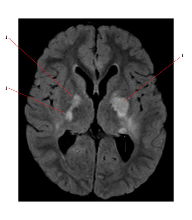
Below is a MR (axial flair image) that demonstrates bilateral signal changes of uncertain etiology (#1 point to these areas) commonly seen in pediatric patients with NF1.

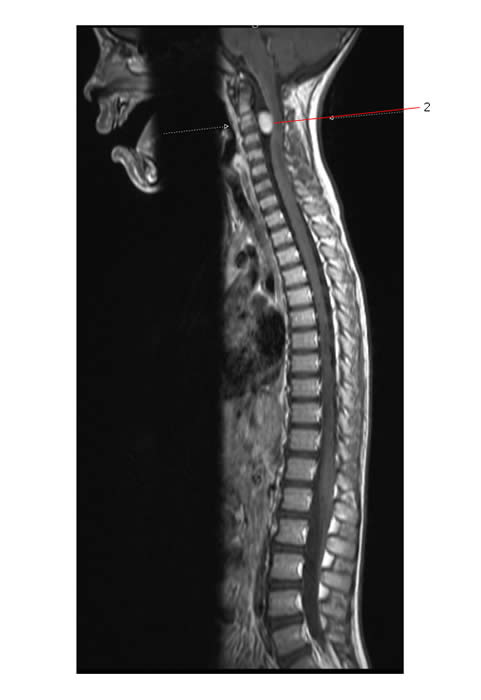
Below is a MR (Sag Postcontrast T1 image) demonstrating a nerve sheath tumour (#2). This is likely a neurofibroma within the vertebral canal, located anterior to and compressing the spinal cord.

Resources:
Neurofibromatosis type 1 (NF1): Atlas of Genetics and Cytogenetics in Oncology and Haematology
Neurofibromatosis at DermAtlas


