Macroscopic Appearance:
The excised primary, untreated is a grey-white fleshy mass.
After chemotherapy, macroscopic evidence of repair may be obvious with:
- Bony widening and sclerosis
- Hemorrhage
- Areas of necrosis
- Cystic degeneration
The amount of necrosis after chemotherapy is of prognostic importance.
Microscopic Appearance:
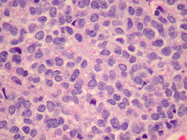
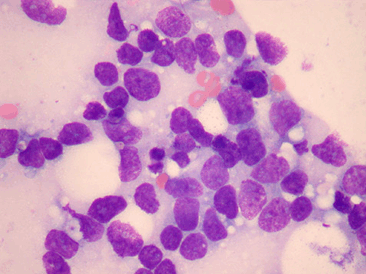
Cytology usually shows:
- Small cells with central, round to oval monotonous nuclei, finely dispersed chromatin and inconspicuous nuclei.
- Cells display a narrow rim of clear or faintly eosinophilic cytoplasm with abundant glycogen [PAS positive].
- There is scant stroma between the cells.
Growth Pattern:
A diffuse growth pattern of wide sheets of cells is common. Other patterns include nests or thick ribbons of tumor cells separated by fibrous septa, organoid patterns that include ‘pseudo-rosettes’ and a filigree pattern.

Ultrastructure:
- ES is comprised of primitive cells with no differentiating features.
- Small round cells:
- Small round hyperchromatic nuclei
- Few organelles are present
- Cell surface simple with cells in close apposition and small intercellular attachments. No basal lamina, and rare primitive adhesion areas [junctions]
- Cells are homogenous
- Abundant cytoplasmic glycogen, typically adjacent to the nucleus [‘cap’]
- Rare dense core granules may be identified
- No extracellular matrix
- Low mitotic rate
ES cells have pale foamy cytoplasm with abundant glycogen:

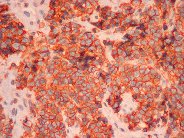
ES cells are positive for CD99:

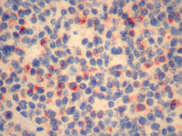
ES cells are positive for synaptophysin:

Typical vs. Atypical Pathologic Features in Ewing sarcoma:
|
Typical |
Atypical |
Cell regularity |
Cellular homogeneity |
Cellular pleomorphism |
Size |
Small |
Large |
Shape |
Round |
Irregular |
Cell layout |
Sheet-like pattern |
Lobular, alveolar, organoid |
Intercellular material |
None |
Collagen bundles |
Intercellular attachments |
Small |
Well developed |
Extracellular matrix |
None |
Microgranular, amorphous |
Cytoplasmic organelles |
None except for lipid droplets, polyribosomes, occasional mitochondria, and pools of glycogen |
Elaborate |
Mitotic rate |
Low |
High |
Cytoplasmic collagen |
Large amount |
Large amount |
Cell proximity |
Very close |
Spaced apart |

