In Ewing sarcoma (ES) magnetic Resonance (MR) imaging is the most effective technique for showing intramedullary and soft tissue extent of disease:
|
MR |
Role |
Shows:
|
Appearance |
|
Limitations |
|
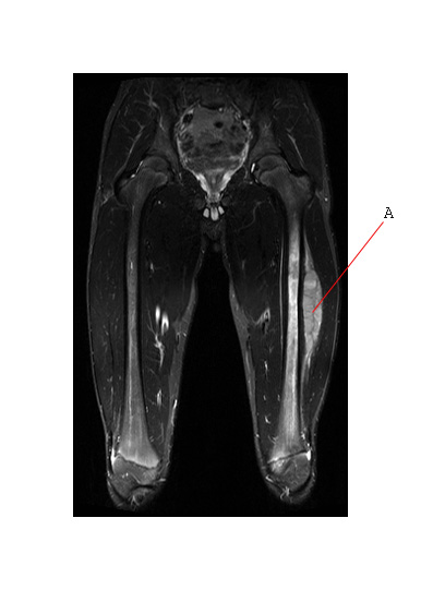
The coronal MR image below is a STIR sequence and shows a large soft tissue mass (A) associated with ES of the femur. This is the same tumor shown in plain films .
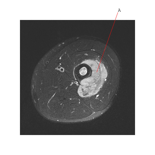
The axial MR image (T2 sequence with added fat suppression) below shows the same soft tissue mass associated with ES (A) arising from the femur.
The MR below shows a soft tissue mass arising from the head of the clavicle (#1) in a patient with ES.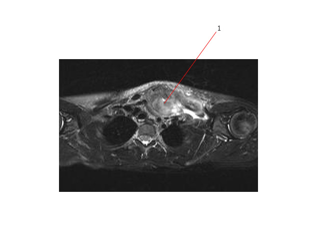
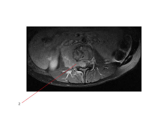
MR below shows ES arising from a vertebral body of the thoracic spine - associated with a soft tissue mass invading into spinal canal (#2) and causing spinal cord compression.
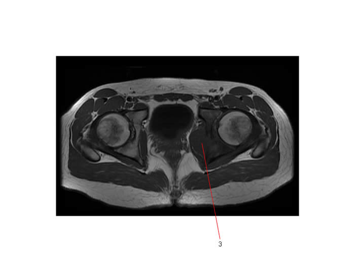
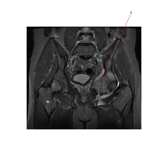
Below are axial and coronal MR scans of ES arising from acetabulum with associated soft tissue mass (#s 3 and 4).
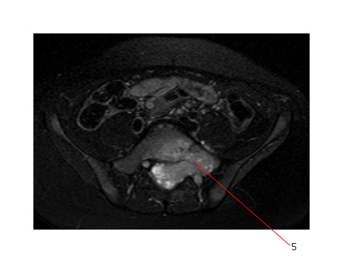
Below is an axial T2-weighted MR image showing increased signal within the right side of S1 and the large intraspinal soft tissue mass which extends through the left S1 neural foramen. This is a sacral ES and #5 points to the epicenter of the tumor,
