CT local
- Unnecessary in the majority of cases
- Some anatomically complex areas may need CT scans for surgical planning
CT chest
- More sensitive than a chest X-ray – detects nodules down to 1-2mm.
- Can have false-positives, especially on high-resolution
- Lungs are the most common site for metastases.
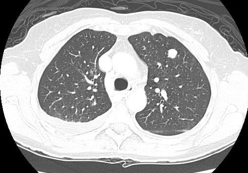
Below is a CT scan which shows a round pulmonary metastatic deposit in the lung that is on your right. There is also a small effusion posteriorly involving the opposite lung.
MR scan
Should be done prior to biopsy so that any resulting edema does not interfere with image interpretation.
Referral should not be delayed while an MR scan is obtained. Subspecialist centres have rapid access to high resolution MR scans and will perform the sequences and series they require.
The information gained from an MR scan is
- Intraosseous tumor extent and evidence of skip lesions
- Relationship of the tumour mass to the neurovascular bundle.
- Can narrow the differential diagnosis in some circumstances depending on the imaging characteristics.
- Dynamic MR scan post Gd-DTPA correlates with histologic response.
MR scans are repeated following the completion of the preoperative chemotherapy to allow response assessment and plan limb surgery.
Examples:
1. The MR below shows extensive lytic bony destruction (#2) and associated soft tissue mass (#1) centred on the distal femoral meta-diaphysis.
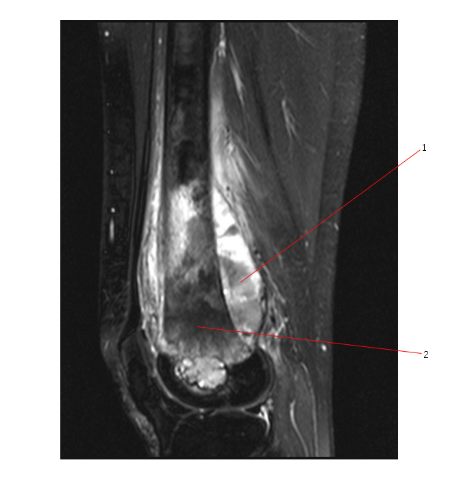
The MR below shows the same tumor.
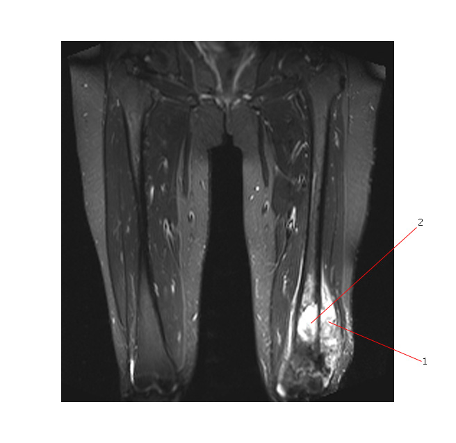
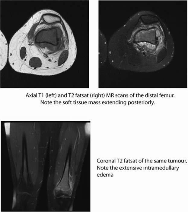
2. MR images of a different osteogenic sarcoma involving the distal femur.
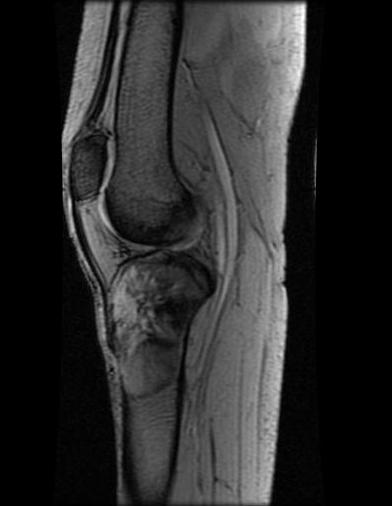
3. MR below shows an OS arising in the proximal tibia and extending to the articular surface of the tibia.
