Bone Marrow Aspirate:

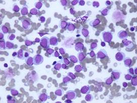
The bone marrow aspirate in AML (above) is
- Hypercellular with a total cellularity of nearly 100%. Approximately 82% of the nucleated cells are blasts resembling those seen in the peripheral blood.
- These blasts are:
- Medium to large in size
- Variably abundant cytoplasm
- No Auer rods
- Mildly irregular nuclear margins and very fine chromatin
- Mature neutrophils show the same dysplastic nuclear morphology as was noted in the peripheral circulation.
- Erythropoiesis is generally normoblastic.
- Megakaryocytes are also generally unremarkable, although rare examples of hypo-lobated forms are identified.
- The sample was stained with non-specific esterase (NSE), myeloperoxidase (MPO) and iron (results are not provided). NSE and MPO staining are both negative, though stainable iron is present within granules.
Bone Marrow biopsy:
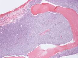
Low power View

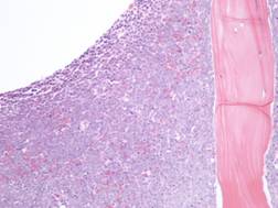
Medium power view

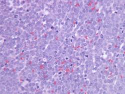
High power View

The bone marrow biopsy above is characteristic of AML and shows that:
- The space is nearly completely occupied by a sheet of blasts.
- Blasts have a limited amount of cytoplasm, generally regular nuclear contours, fine chromatin, and prominent nucleoli.
- Residual hematopoiesis is minimal.

