Location
Most Wilms tumors are unicentric lesions, but may arise multifocally in the kidney.
- 5% involve both kidneys at presentation or subsequently
- 10% are multicentric unilateral tumors (within the same kidney)
Wilms may arise anywhere in the kidney.
Extrarenal Wilms tumors
- Sometimes Wilms tumors may arise from outside the kidney
- Rare
- Generally occur in the retroperitoneum adjacent to the kidney.
- Origins thought to include displaced microscopic elements or extragonadal germ cells.
Spread
Local:
- Local spread initially involves the renal sinus or intrarenal blood and lymphatic vessels and is intra-renal.
- Penetration through the renal capsule (extrarenal spread) often occurs next. Tissue or vascular invasion, or both, may be seen.
- Tumor cells may invade into the perirenal fat. Tumor invasion into this area is often associated with an inflammatory or granulation tissue response in the perirenal connective tissue. Sometimes it is very hard to demonstrate tumor cells. This is called the “inflammatory pseudocapsule” (IPC). There is often associated hemorrhage into perirenal tissues or extensive necrosis of tumor just beneath the capsule. The presence of IPC is currently acceptable for stage I if there is no capsular penetration by tumor cells.
- Four features related to the extension of tumor within the kidney are associated with an increased risk of relapse in Stage I Wilms Tumor. These are:
- Presence of an inflammatory pseudocapsule
- Extensive infiltration of the renal capsule
- Involvement of the renal sinus
- Tumor in the intrarenal vessels
- The renal hilus is not usually encapsulated; therefore hilar invasion has not been sufficient grounds for stage II designation. However, extensive hilar invasion is associated with aggressive tumor behavior, and previous NTWS staging has been based on extension of tumor past the hilar plane. The hilar plane is an anatomic landmark formed by the 2 medial-most limits of the kidney. This landmark is hard to assess in practice because of the anatomic distortion caused by bulging tumors. In the newest staging system vascular invasion outside of the kidney substance is considered grounds for stage II designation.
Lymphatic:
- Regional nodes (include the renal hilar nodes or the para-aortic nodes) may be involved.
Distant:
Most common sites of distant spread are:
- Lungs (occur in about 15% at presentation)
- Liver
- Other metastatic sites are uncommon (brain metastases can occur very occasionally in FH Wilms)
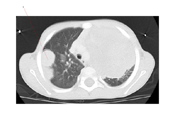
The CT below shows pulmonary metastases from Wilms tumor. #1 points to a large area of nodular disease.

Bone metastases are a hallmark of clear cell sarcomas of kidney (CCSK).
Both CCSK and rhabdoid tumor are associated with brain metastases.

