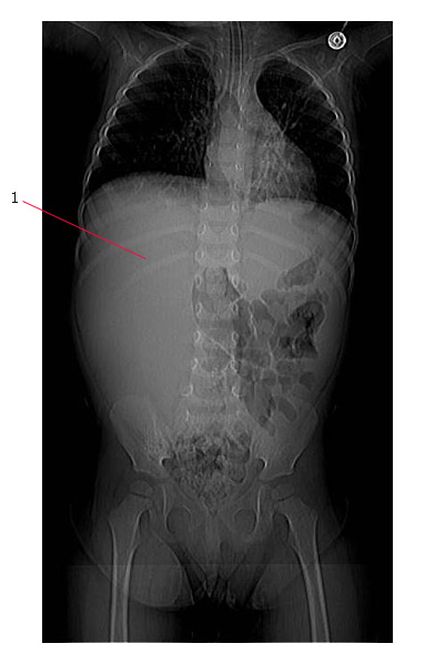
The plain abdominal film below shows a large, unilateral abdominal mass (#1) displacing bowel (no bowel gas can be seen on the right side of the abdomen). This 3 year old child had a Wilms tumor.
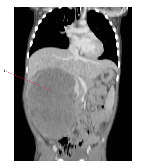
Here is a coronal CT scan which shows the same tumor (#1).
Imaging is used to evaluate the extent of both local and distant disease
Local disease
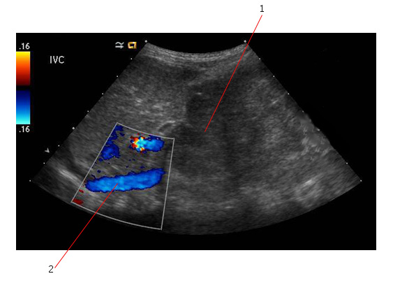
Ultrasound scan is usually the first investigation to confirm the presence of an abdominal mass. This will also give information about the presence of tumor thrombus in blood vessels.
In the ultrasound scan below #1 points to the tumor and #2 to the inferior vena cava (IVC) which is patent.
CT scan is an especially important imaging study pre-operatively to establish:
- Disease extension to involve adjacent organs
- Vascular extension
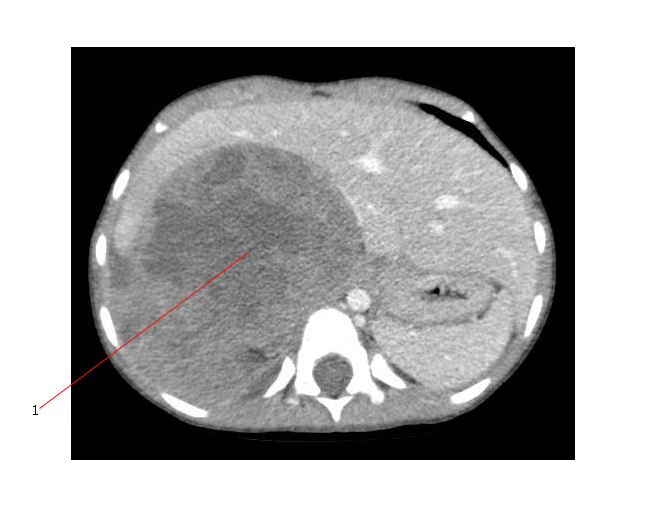
The axial CT scan below shows a large, heterogeneous Wilms tumor displacing the liver. #1 points to the necrotic center of the tumor.
Distant disease:
- Metastatic disease can involve the lung and less frequently, the liver.
- Lymph nodes at the renal hilum may be involved.
- The contralateral kidney is sometimes also affected by disease (Stage V - Bilateral tumor).
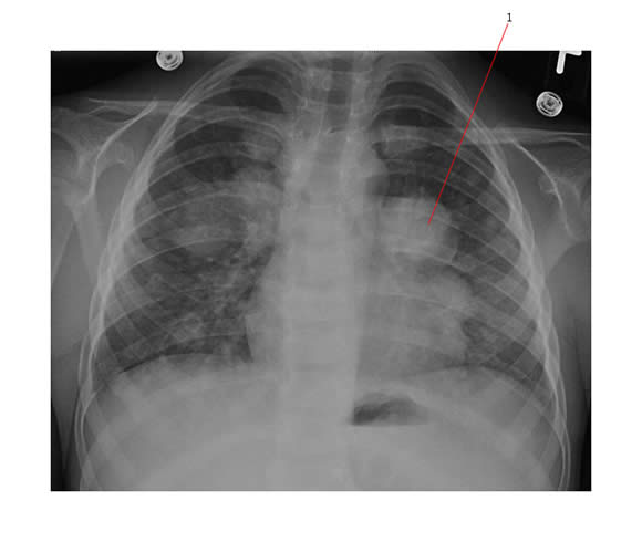
The chest X-ray below shows multiple pulmonary metastases from Wilms tumor (#1 points to a particularly large nodule).
CT scan of the abdomen and chest is the most accurate investigation for both local and distant staging.
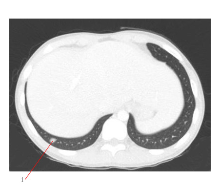
The CT scan below shows a small pulmonary nodule (#1) at the lung base. This nodule of metastatic Wilms could only be detected by CT scan.
Radiology findings in Wilms Tumor:
Abdominal Ultrasound |
CT scan |
|
Role |
Establish presence of renal mass
Identify
|
Further evaluate size, nature and extent of mass:
Evaluate
|
Appearance |
Often fairly homogeneous with overall echogenecity similar to liver
Increased or decreased echogenic foci from hemorrhage, necrosis
May contain multiple fluid pockets (cystic degeneration) | Heterogeneously enhancing mass
Lower in attenuation than surrounding normal renal parenchyma on contrast enhanced CT (due to hemorrhage or tumor necrosis)
Very rarely may be foci of high attenuation within mass indication calcification. |
Link:


