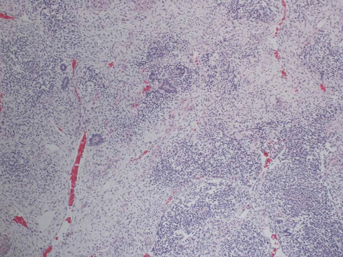
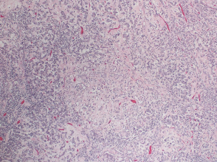
Wilms tumor (WT) is an abnormal proliferation of the mesanephric blastema with no differentiation into tubules or glomeruli and usually arises from three tissue elements:-
- Blastema – microscopically undifferentiated small round blue cells arranged in a diffuse or organoid pattern
- Epithelium - epithelial structures [tubules or glomeruli] develop out of condensations of blastema and may simulate the nephrogenic development of the kidney, however larger capillary formations unlike normal developing kidney may also be present while heterologous squamous or mucinous elements may also be identified
- Stroma - this is usually manifest as immature spindled cells, but heterologous differentiation may result in the presence of skeletal muscle, osteoid, or fat.
Tumors with all three components are termed triphasic, but some may be biphasic or monophasic only [e.g. Blastemal WT], whereas tumors with a majority of heterologous elements are be termed ‘teratoid’.
Tumor histopathology is very important and affects prognosis. Tumors are categorized as having either favorable (FH) or unfavorable histology (UH):
Favorable versus Unfavorable Histology in Wilms Tumor:
| Favorable Histology (FH) | Unfavorable Histology (UH) |
|
| Features |
|
Extreme anaplasia 3 cytological abnormalities:
Anaplasia may be diffuse or focal; even in one or two foci is enough to impart a markedly worse prognosis
These changes are associated with high rates of relapse and death |
| Proportion of tumors |
|
|
Gross examination
- Any part of the kidney may be involved.
- The tumor is usually unilateral, but to 11% may be multifocal, and 5-10% of cases may be bilateral.
- Macroscopically the tumor is well demarcated, solid or cystic and often surrounded by a psuedocapsule of compressed, atrophic renal tissues.
- The tumor cut surface is variegated and may be firm and bosselated, necrotic, myxomatous, or cystic.
- Invasion of vessels and ureter may occur.
- Regional lymph nodes may be involved by tumor, but enlarged draining nodes may also reflect reactive sinus histiocytosis.
- Extra renal Wilms are very rare.
Microscopic examination
There are several histologic classifications for Wilms tumor.
Histological Characteristics of Wilms Variants:
Classic triphasic FH Wilms tumor |
|
Blastemal Predominant |
|
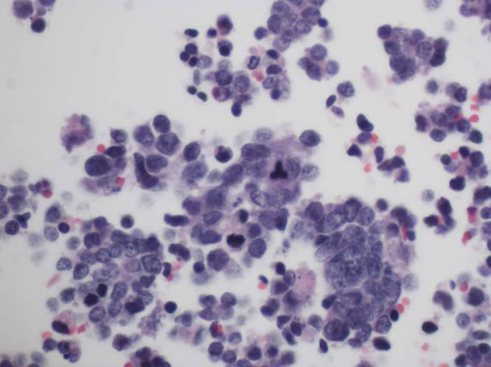
Anaplastic Wilms tumor |
|
Pathologic appearance of Wilms tumor:
Classic triphasic FH Wilms tumor |
|
More differentiated FH Wilms |
|
Anaplastic Wilms tumor with diffuse anaplasia - pleomorphic hyperchromatic nuclei and abnormal mitoses. |
|
Unfavorable Histology (UH)
- Histologic feature of unfavorable prognosis is the presence of anaplasia.
- Anaplasia = extreme cytological atypia.
- Anaplasia may be present in any of the three cell types in WT and is defined as the presence of 3 histologic criteria:
- Large nucleus [nuclei with a diameter 3 times that of an RBC in one dimension and 2 times that of an RBC in another dimension] (or the older definition of nuclear size three times that of neighboring cells of same histologic type)
- Hyperchromasia of the enlarged nuclei
- Atypical [multipolar] mitoses
- All three features must be present to establish a diagnosis of UH.
- Anaplasia is rare in WTs from children <2 years of age but there is an increased incidence thereafter until 5 years of age (13% of cases).
- The overall incidence in all cases of WT is 5%.
- Anaplasia is associated with greater resistance to chemotherapy as opposed to a more aggressive growth pattern.
- Anaplasia may be focal or diffuse. It is defined as diffuse when it is:
- present outside the kidney
- identified in a random biopsy
- present in a background of severe nuclear atypia
- present in multiple slides whose precise location cannot be determined
Stages 2-4 WT - those patients with focal anaplasia have the same outcomes to that of Favorable Histology WT, while the outcomes for those with diffuse anaplasia is poor.
Nephrogenic rests (NR)
- Foci of abnormally persistent embryonal tissue are recognized precursors to WT.
- NRs found in the kidneys of almost 1% of unselected pediatric autopsies
- Present in up to 35% of kidneys which develop unilateral WT and almost 100% of kidneys involved by bilateral WT.
- The incidence of NR is far in excess than that of WT - so only a small proportion of NR develop into WT.
- The finding of multiple or diffuse NRs in a kidney bearing WT indicates an increased risk for development of WT in the opposite kidney, a factor to be considered in planning clinical follow-up.
NR are classified by their position within the renal lobe.
- Intralobar nephrogenic rests [ILNR]
- Tend to occur deep within the lobe
- Reflect an earlier developmental insult to the kidney
- Usually stroma rich and infiltrate surrounding renal parenchyma
- More strongly associated with the WAGR and Denys-Drash syndromes, and the development of a stroma rich WT at an earlier age
- Perilobar nephrogenic rests [PLNR]
- Located at the lobar periphery
- Reflect later developmental disturbances in nephrogenisis
- Comprise blastema and tubules sharply demarcated from adjacent normal kidney
- Associated with BWS and the development of epithelial or blastemal predominant tumors
Links


