
RT Beam Basics
Beam characteristics - Penumbra
Penumbra
- The rapid decrease at the edges of the radiation beam is called the penumbra region
- Usually defined as the space between the 80% and 20% isodose lines.
Often, the 50% isodose line (defined as the geometric penumbra) is the standard for delineating the effective area of the beam.
The light field that comes out of the linear accelerator indicates where the 50% isodose line will be.
The size of the penumbra region is dependent on many factors such as:
- source to skin distance (SSD)
- source size
- transmission through the jaws of the machine
- energy
As we rarely deal with sources anymore, let’s just talk about the factors we may encounter:
SSD (Source to Skin Distance)
Increasing the SSD will cause the penumbra to increase in size as the photon particles have more opportunity to veer off course slightly and increase the penumbra.
Energy
On the other hand, increasing energy has a tendency to sharpen the penumbra. As the energy of the radiation beam increases, there is less side scatter of the beam so the penumbra region is smaller.
However, with most modern energy beams (6, 10 and 18MV), there is relatively little difference in the penumbra region.
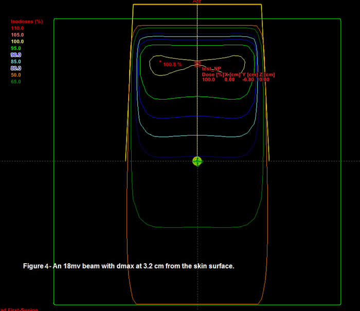
Figures 3 and 4 below show the penumbra region of beams of different energy. You can see the 4MV and 18MV beams look relatively similar in the penumbra region.
Figure 3
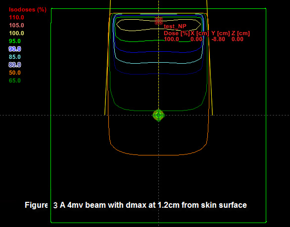
The clinical effect of the larger penumbra regions of extended SSD’s or low energy beams is a lack of sharpness at the edge of the field which can be important for junctioning fields or when the beam is near critical structures.
Figure 4
Dmax
When a radiation beam interacts with tissue, a certain depth of tissue is required before we see the maximum amount of radiation (maximum dose or dmax). This region is called the build-up region and is responsible for the skin sparing effect.
The dose at dmax is defined as 100% and then the radiation dose decreases as the depth increases, the energy being absorbed within the tissue.
The depth of dmax and thus the size of the build up region is energy dependent. Figure 3 and 4 show the point of dmax for 4MV and 18MV energies. You will quickly see the different penetration for each beam.
For the record, the depths of dmax for some common energy’s are:
4MV: 1.2cm
6MV: 1.5cm
10MV: 2.5cm
18MV: 3.2cm