Superior Vena Caval (SVC) Obstruction
and
Superior Mediastinal Syndrome (Central Airways Obstruction)
Superior vena caval syndrome is when there is obstruction of the superior vena cava by a mediastinal mass. Superior mediastinal syndrome usually refers to other structures being involved such as the trachea.
Respiratory distress is often the presenting symptom for children with a thoracic malignancy.
A mass in the anterior superior mediastinum can cause:
- Superior vena cava syndrome (SVCS)
- Superior mediastinal syndrome (SMS)
The SVC is a thin walled vessel which is easily compressed by tumors or infection arising in adjacent lymph nodes and thymus.
SVCS is caused by compression, obstruction or thrombosis of superior vena cava.
Different causes of SVC syndrome in children:
Bold lettering shows commonest causes
| Congenital | Thrombotic complications after surgery for congenital heart disease. | |
Inflammatory |
Infection | TB Histoplasmosis |
| Non-Infective | granulomas | |
| Venous thrombosis after line insertion | ||
Neoplastic |
Benign | Hemangioendothelioma |
| Teratoma | ||
| Thymoma | ||
| Malignant | T cell lymphoblastic lymphoma Non Hodgkin Lymphoma commonest cause (70%) |
|
| Acute T cell lymphoblastic leukemia | ||
| Hodgkin lymphoma | ||
| Neuroblastoma | ||
| Sarcoma | ||
| Thyroid carcinoma |
In up to 70% of children with SVC Syndrome there is some airway compression in mediastinum.
Symptoms and Signs
- Dyspnea (short of breath) and orthopnea (can't lie flat - so have to sleep with extra pillows)
- Cough and wheezing
- Dysphagia (difficulty swallowing)
- Facial swelling and conjunctival suffusion
- Venous distention in upper body
- Headache, anxiety, confusion, syncope
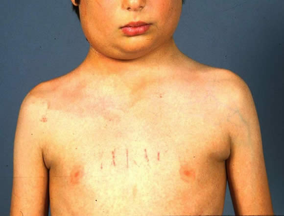
The image below shows a child with SVC compression. He has facial swelling and venous distention involving the upper body. He also has right sided cervical lymphadenopathy.

Investigation
- Chest X-ray
- CT scan chest
- Echocardiogram
- Blood work - CBC, LFTs, clotting studies.
- Anesthesia consult
- PFTs
- Biopsy if possible.
- Try to find other areas to approach for biopsy such as bone marrow, peripheral lymph node, tap pleural or pericardial effusion.
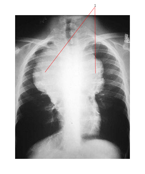
The chest X-ray below shows a massive anterior mediastinal mass (#1) due to T cell lymphoblastic lymphoma.

Therapy
A chest X-ray should be done before transport of any child with possible malignancy, especially those who have a hematologic malignancy.
The first steps are to:
- Sit child upright
- Reduce anxiety in child and keep child calm if possible
- Steroids may be required if severe airway concerns (if treating then need to manage tumor lysis risks)
- Often impossible to intubate these kids and need to involve anaesthesia for airway management
- Be wary of giving any sedation as this may exacerbate the airway compromise.
- Emergency measures to maintain oxygenation - Oxygen by mask or nasal prongs
In hospital:
- Dexamethasone and supportive care in ICU.
- Radiation Therapy:
- Effective for Non-Hodgkin lymphoma (NHL) often within 12 hours.
- May make pathology impossible to assess after.
- Chemotherapy
- Child may have other problems that need systemic therapy such as hyperleukocytosis.
- Increasingly used with systemic steroids.
- Surgery
- May be necessary if the tumor is not radiosensitive or chemosenstive. Example would be teratoma.

