They are very often embryonal tumors (20% are botyroid).
Bladder tumors
- These tumors tend to grow intraluminally before invading bladder wall, in or near the trigone and look polypoid.
- Usually occur in children under 4 years old.
- There may be haematuria, urinary obstruction and occasionally the passage of tissue per urethra.
- Bladder tumors tend to remain localized, but may invade the prostate.
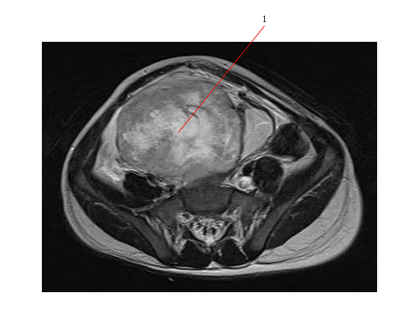
The MR below shows an embryonal RMS (#1) which has arisen from the dome of the bladder:
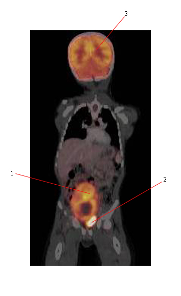
Below is a PET scan of the same patient showing the RMS (#1) arising from the displaced bladder (#2) and normal uptake in the brain (#3).
Prostate tumors
- Prostate tumors produce large pelvic masses, sometimes with urethral strangury and constipation.
- Tend to occur in infants and in older age groups.
- Prostate tumors have a greater tendency to infiltrate adjacent tissues (than bladder).
- Often spread early to distant sites such as the lungs and less frequently to bone.
- Prognosis is worse than for bladder.
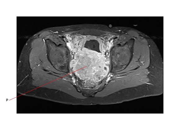
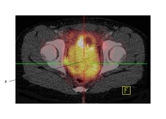
The axial MR image below shows a large prostatic RMS (P) arising in the pelvis of a 9 year old boy.
Below is a PET scan image of the same tumor, showing FDG avidity.
Vaginal tumors
- Usually botryoid (look grape like in appearance) and are almost always found in very young children (2 to 3 years old).
- Cervical and uterine sarcomas are rare and tend to occur in older children and usually present with a mass and /or discharge.
- Vaginal RMS is far more common than cervix or fundus.
Paratesticular tumors
- Usually produce painless unilateral scrotal or inguinal enlargement.
- Intrascrotal RMS usually arises from the spermatic cord.
- Retroperitoneal nodes are often involved.
- Orchiectomy should be performed through an inguinal incision, with high ligation of the spermatic cord (the spermatic cord is resected at the level of the internal inguinal ring). Scrotal skin is only resected if it is involved. Hemiscrotectomy can be performed if there is residual disease in the scrotum and this is preferred to routine scrotal RT.
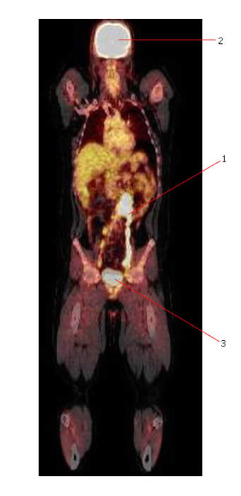
The PET-CT scan below shows avid FDG uptake in extensively involved retroperitoneal nodes (#1) from paratesticular RMS. #2 points to normal brain. #3 points to normal bladder.
