Location
It is common for craniopharyngiomas to involve the sella and the suprasellar space, but tumor location, size, texture, and adherence to surrounding structures may vary 4,8.
- 70% of tumors involve both the infrasellar and suprasellar regions.
- Of those isolated to one particular region, 20% are restricted to the suprasellar region, while 10% are solely infrasellar.
- Tumors may also be prechiasmal or retrochiasmal.
- Prechiasmic:
- 30% of childhood craniopharyngiomas are prechiasmic.
- Tumors originate between the optic nerves and push them posteriorly.
- Tumors are generally more accessible and less adherent to vital suprasellar structures.
- Retrochiasmic:
- 70% of childhood craniopharyngiomas have a retrochiasmic component.
- Tumors usually extend superiorly.
- Retrochiasmic lesions may grow into the 3rd ventricle, producing hydrocephalus, compress the optic tracts, or grow into the hypothalamus.
- If the tumor is multicystic, there may be a cystic extension into the basal cisterns and the posterior fossa.
Tumor Growth
These tumors are benign and grow slowly, but:
- Craniopharyngiomas can grow to reach an enormous size (because of an enlarging cystic component) causing significant neurological impairment.
- While often labeled as “encapsulated”, this term is often debated due to the infiltrative nature of the neoplasm.
- Craniopharyngiomas have been known to infiltrate the brain, the tuber cinereum, and the hypothalamus.
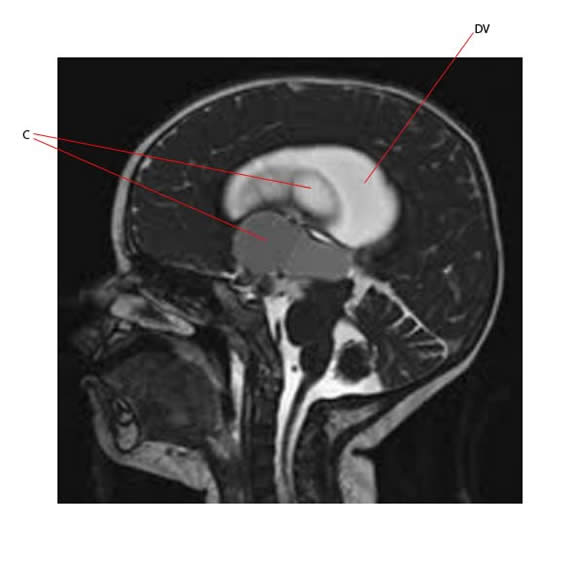
The MR of a 4 year old boy below shows large cysts (C) associated with a craniopharyngioma. The tumor is arising from the suprasellar region. Cystic masses indent and fill the third ventricle causing obstructive hydrocephalus and dilated ventricles (DV).


