Cellular Origin:
Pediatric craniopharyngiomas arise from cellular remnants of the Rathke pouch4,12
Possible histological origins of these tumors may be:
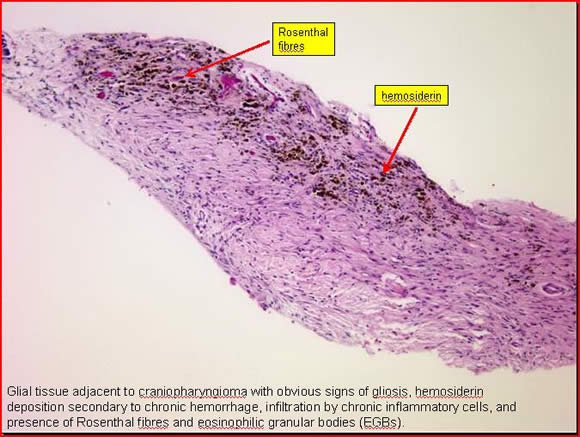
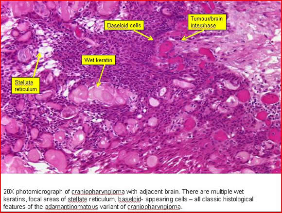
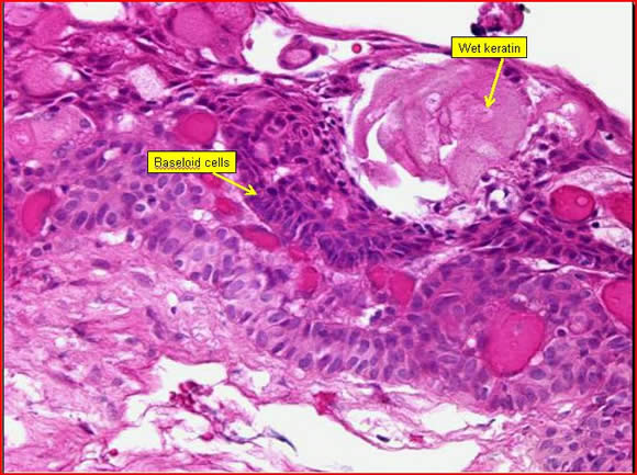
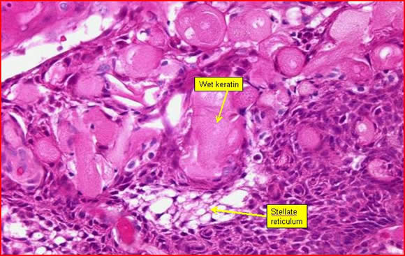
- Embryonic cell rests of enamel organs located adjacent to the tuber cinereum along the pituitary stalk (most widely accepted theory) – adamantinomatous variant
- Cellular remnants of Rathke’s cleft – papillary variant
- Metaplasia in cells of the adenohypophysis (an alternative to derivation from embryonic cell rests)
Craniopharyngiomas are classified into three histologic types4,13,17
- Adamantinomatous
- Papillary
- Mixed
Intracranial Location and Gross Pathology:
- Predominantly appears as calcified cystic suprasellar mass which produces visual and pituitary defects
- May also appear in other locations such as nasal, 3rd ventricular, pineal, and infratentorial
- Rupture of cyst and leakage of content can cause recurrent asceptic meningitis or Mollaret’s meningitis
- Often associated with tenacious adherence to adjacent normal vascular and neural structures which makes complete surgical resection challenging.
Characteristics:
Adamantinomatous |
|
Papillary |
|
Mixed |
|
Summary of histological properties of craniopharyngioma:
Location |
|
Macroscopic appearance |
|
Microscopic appearance |
|
Benign/Malignant |
|
Calcification |
|
Colour |
|
Size |
|
Texture |
|
Keratin pearls |
|
Carcinomatous areas |
|


