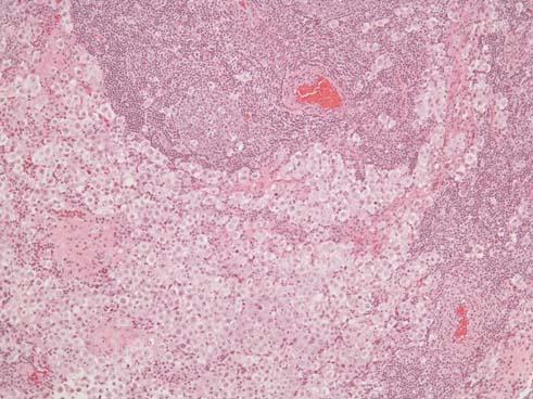
The slide below: Germinoma cells with lymphoid aggregates and lymphocytes infiltrating fibrovascular septae (H and E x 100).
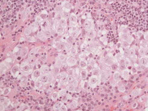
The slide below is a higher power of germinoma cells.
- Germinoma cells have:
- discrete cell membranes
- clear cytoplasm
- vesicular nuclei
- prominent nucleoli
Mitotic activity is brisk within this tumor (H and E x400).
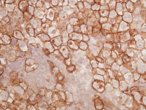
The slide below: Germinoma cells showing membranous and golgi staining with c-kit protein (c-kit immunohistochemical stain x400).
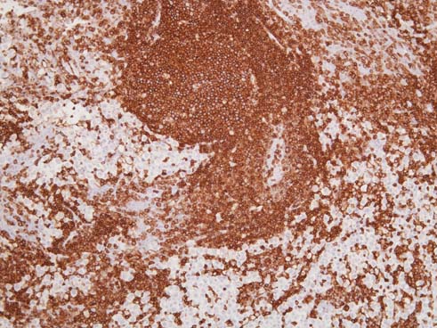
The slide below: Leukocyte common antigen stain showing staining of lymphocytes in lymphoid aggregates, in fibrovascular septae and scattered within the tumor (LCA immunohistochemical stain x 400).