Local imaging
The tumor should be imaged prior to any biopsy to accurately assess the extent of disease.
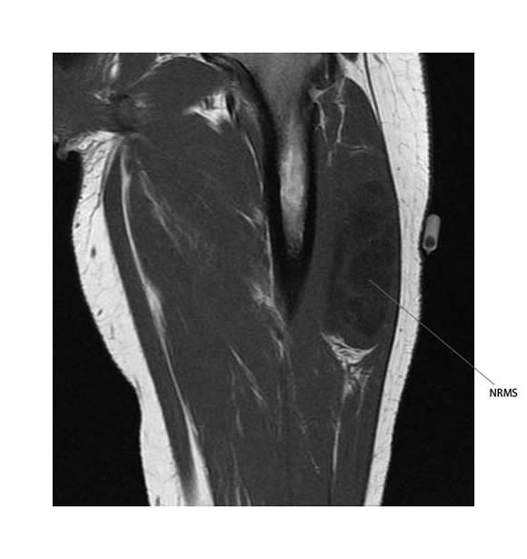
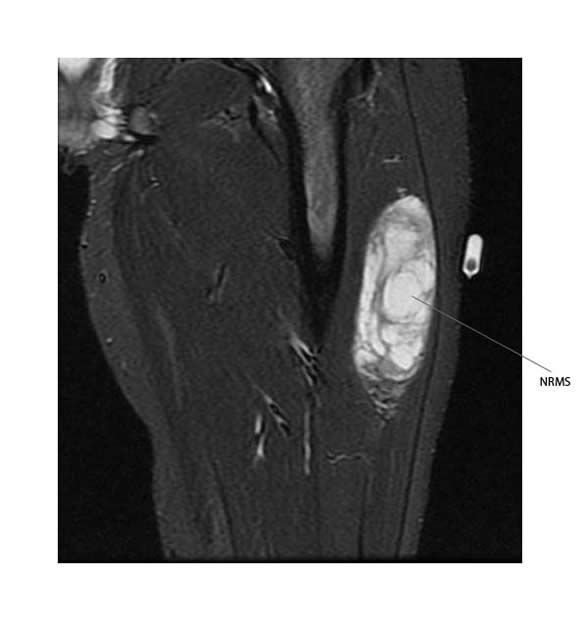
MR scan
- Best imaging modality to assess the extent of a soft tissue sarcoma
- Primary tumor is typically:
- Well circumscribed mass within or adjacent to muscle
- Assess:
- Diameter of tumor
- Relationship to neurovascular bundle
- Characteristics
- T1
- Intermediate to low signal intensity, similar to adjacent muscle
- Heterogenous if hemorrhage, calcification, necrosis and myxoid material present
- Enhancement of solid components with gadolinium
- T2
- Intermediate to high signal intensity
- Heterogenous if hemorrhage, calcification, necrosis and myxoid material present
T1 sequence:

T2 sequence:

For intra-abdominal retroperitoneal and intrathoracic sarcomas, CT scan may give accurate information about the local extent of disease.
Distant metastatic disease
CT scan
- To exclude distant metastatic disease in lungs
- To assess possible metastatic disease to liver and abdomen for liposarcoma
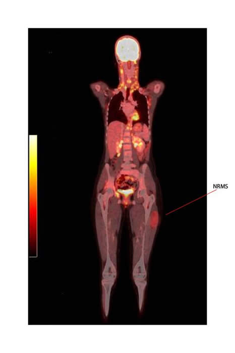
CT-PET scan
- FDG positron emission tomography has not been used routinely in the assessment of follow up of pediatric non-RMS.
- There is evidence in adult tumors that this modality can be used as part of initial staging.
- CT-PET allows high sensitivity for the detection of various sarcomas and accurate discrimination between newly diagnosed low-grade and high-grade sarcomas.
- In one series over 90% of adult soft tissue sarcoma were FDG avid, but PET-CT results rarely changed managment.

The PET-CT above shows increased uptake in a non-RMS of the thigh.
References and Resources:
FDG PET/CT in Initial Staging of Adult Soft-Tissue Sarcoma. Sarcoma Volume 2012 (2012), Article ID 960194,
FDG PET/CT imaging in primary osseous and soft tissue sarcomas: a retrospective review of 212 cases.
Eur J Nucl Med Mol Imaging. 2009 Dec;36(12):1944-51. doi: 10.1007/s00259-009-1203-0.

