Plain films:
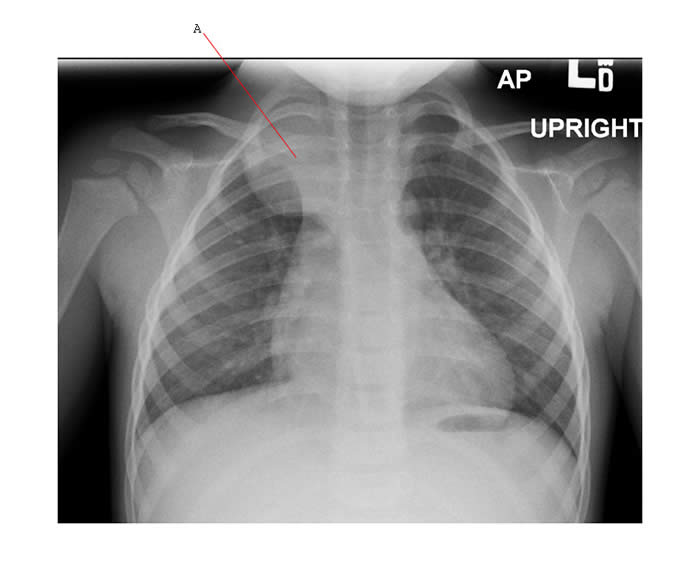
Below is a chest X-ray showing a mediastinal mass (A) - this was a thoracic neuroblastoma.
CT scan:
- Preferred for posterior mediastinal tumors.
- MR may be preferred to evaluate paraspinal tumors.
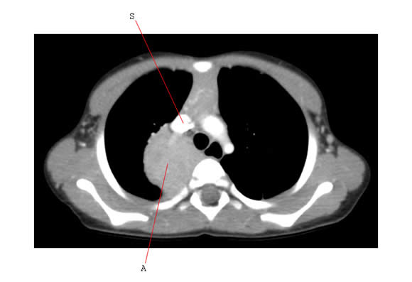
The CT scan below is of the posterior mediastinal neuroblastoma shown on plain film (A). The SVC (S) is visible on this axial image immediately anterior to the mass and is not compressed.
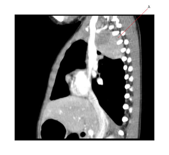
The sagittal image of the same tumor below shows fingers of tumor extending into the intervertebral exit foramina (A) - common for neurogenic tumors in this region to do this.
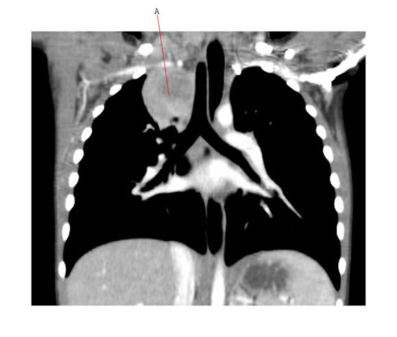
Coronal CT scan cut below showing this intrathoracic neuroblastoma (A) and relationship to the airway.
Abdominal Tumors
CT scan:
- used routinely to evaluate the extent of abdominal and pelvic tumors
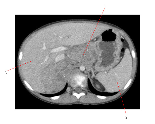
The axial CT scan below shows an intrabdominal neuroblastoma. The mass (#1) contains dystrophic calcification, crosses the midline and is associated with extensive adenopathy. #2 is the spleen and #3 is the liver.
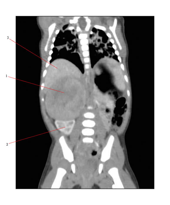
The coronal CT scan below shows a large primary neuroblastoma arising from the adrenal (#1). The kidney (#2) is displaced down and the liver (#3) is pushed up.