Radiology
Radiological investigations:
- Confirm the location of disease
- Are critical in tumor staging to assess
- local extent
- distant spread
Plain chest X-ray is often a useful initial investigation
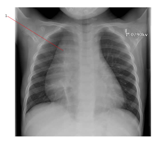
The chest X-ray below was taken on a child who presented with difficulty breathing and a very high WBC count. It shows a right sided mediastinal mass (#1). He had acute T cell lymphoma/leukemia.
Examples of looking at the local extent of Disease
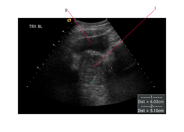
Ultrasound scan can help to delineate abdominal and pelvic tumors.
The transverse ultrasound image of the pelvis below showed a soft tissue mass (#1) deep to the urinary bladder (shown by B). This was a prostatic rhabdomyosarcoma.
Computerized Tomography (CT) scan
CT shows:
- Internal organs
- Tumor
- location and relationship to organs
- size/extent of tumor
- associated bone destruction
Scans often show more if iodinated intravascular contrast media is given.
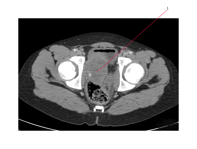
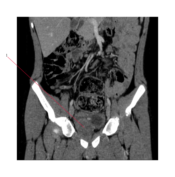
Axial and coronal CT images below show a complex soft tissue mass, arising from the prostate gland (#1). This is the same prostatic rhabdomyosarcoma seen in the ultrasound scan above. A small amount of air is seen in the urinary bladder following placement of a catheter.
Magnetic Resonance Imaging (MR) Scan
This technique has revolutionized imaging of the central nervous system. MR also shows the soft tissue extent of tumors very accurately.
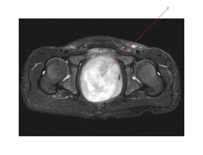
Below is a MR scan of a much larger prostatic rhabdomyosarcoma (#2) than the one previously shown. The soft tissue extent of the tumor can be clearly seen.
Examples of Looking at the Distant Extent of Disease
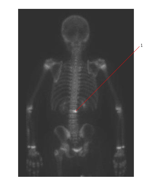
Below is a bone scan on a child with osteogenic sarcoma. There is increased uptake in the body of L2 consistent with bone metastases (#1).
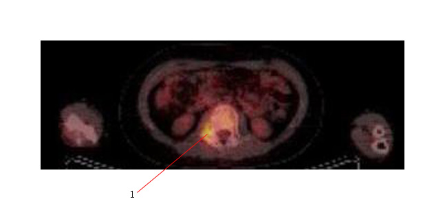
Below is a PET scan of the same child with axial cuts through the same area. The PET shows significantly increased activity in the region of the metastatic deposit (#1).
