Physical Examination
In the examination of a child with possible malignancy, special attention should be paid to the following features :
General health
- Is the child generally well and comfortable / sick and in pain?
- Is the child febrile?
- Is the child pale /anemic?
- Skin
- Is there a rash? - for example purpuric rash in thrombocytopenia
- Are there many bruises?
- Vitals - pulse, BP
Evidence of Congenital abnormality or Syndrome
- Optic gliomas are associated with Neurofibromatosis.
- Wilms tumor is associated with:
- Aniridia
- Beckwith-Weidemann Syndrome and hemi-hypertrophy
- Genito-urinary abnormalities
Assessment of Physical Growth
- Head circumference (standard head circumference chart)
- May be enlarged in an infant with a brain tumor and hydrocephalus.
- Height and weight (percentile charts for girls and boys)
Developmental Assessment
- When did the child achieve their developmental milestones?
- Have any of these skills been lost?
- May see loss of acquired skills with brain tumors.
Examination of Lymph node Groups
All groups should be examined:
- Occipital
- Posterior auricular
- Preauricular
- Tonsillar
- Submandibular
- Submental
- Cervical (anterior - upper and lower, posterior - upper and lower)
- Supraclavicular and infraclavicular
- Axillary
- Epitrochlear
- Popliteal
- Inguinal
Many children have small but enlarged lymph nodes in the cervical, axillary and inguinal regions.
Supraclavicular adenopathy is often due to illness. Left supraclavicular adenopathy is often due to malignancy.
Nodes over 2.5 cm in diameter are pathologic. Malignant lymph nodes are generally firm, rubbery and matted.
Examination of Head and Neck
This includes careful examination for:
- Facial swelling
- Eye signs (see below)
- Ears - is there any bulging of underlying mastoid? Does the tympanic membrane look normal?
- Oropharynx
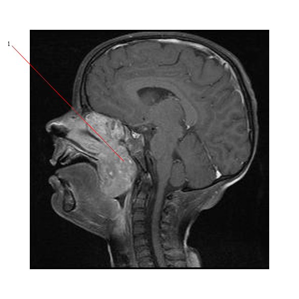
The MR below was done on a child who had a long history of nasal congestion and increasing difficulty breathing at night. He could only breathe through his mouth. He had chronic otitis media. The scan shows a large tumor (#1) filling his nasopharynx and extending inferiorly to also obstruct his oropharynx. This proved to be a rhabdomyosarcoma.

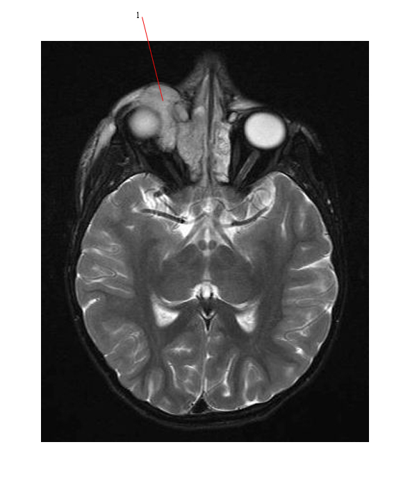
The diagnosis was only suspected when he developed proptosis of one eye as shown below. The tumor (#1) can be seen extending into the orbit. By the time the diagnosis was made, his disease was very advanced.

Examination of Eyes
Many different eye signs can be present in pediatric oncology patients:
- Proptosis - can be due to
- rhabdomyosarcoma (orbital and parameningeal)
- Optic nerve glioma
- Orbital bruising
- periorbital edema can occur in children with metastatic neuroblastoma to the orbit
- called "racoon eyes"
- Horner syndrome - due to involvement of the sympathetic chain in neck or upper chest. Usually seen in neuroblastoma.
- enopthalmos
- anhydrosis
- meiosis
- ptosis
- Papilloedema
- associated with raised intracranial pressure
- Cranial nerve palsies
- VI nerve palsy associated with raised intracranial pressure
- Retinal disease
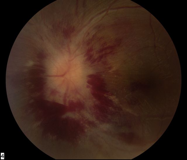
- hemorrhage/exudates secondary to leukemic involvement of the optic nerve head and posterior retina (as shown below)

- Retinal tumor - retinoblastoma
- Strabismus
- Photographs show leukocoria (cat's eye reflex)
- Reduced visual acuity - retinoblastoma, optic nerve/chiasm tumor
Link to overview of Retinoblastoma
Examination of the Cardiovascular System
- Hypertension can occur in:
- Neuroblastoma due to excessive catecholamine secretion or due to renovascular compromise and increased renin levels
- Wilms tumor - due to renin produced by Wilms tumor cells or again due to renovascular compromise.
- Signs of SVC obstruction - usually associated with a large mediastinal mass due to lymphoma / leukemia.
- Signs of a pericardial effusion - again often associated with lymphoma
Examination of the Respiratory system
- Increased respiratory rate due to reduced lung function (effusion or airways obstruction).
- Chest wall swelling and tenderness due to underlying tumor
- Reduced air entry:
- pleural effusion secondary to involvement of the pleural cavity by a tumor such as Ewings sarcoma
- collapse/consolidation of underlying lung due to tumor
Examination of the Abdomen
- Find out if there is any abdominal pain and ask the patient/parents to identify painful area if possible before any examination begins.
- Inspection for visible swelling of abdomen
- Often found in Wilms tumor
- Tenderness
- Palpable organomegaly
- splenomegaly may be present in lymphoma
- Palpable mass
- may be the presentation of a rhabdomyosarcoma
- Bowel sounds present? normal? (subacute bowel obstruction can be a presentation of Non-Hodgkins lymphoma)
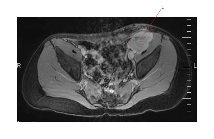
The MR below was done on a child who complained of low abdominal pain and on examination was found to have a tender mass palpable in the left iliac fossa. This scan shows a T2 bright lobulated mass (#1) which proved to be a rhabdomyosarcoma.

Examination of the Nervous system
- Level of alertness
- may be reduced in patients with raised ICP
- Cranial nerve abnormalities and papilledema
- May be associated with raised ICP
- The brain tumor itself may cause cranial nerve palsies (eg. brain stem glioma)
- Motor strength
- Brain tumors can affect the motor cortex and cause hemiplegia
- Spinal cord tumors can cause paraplegia.
- Sensory deficits
- Cerebellar dysfunction
- Common at presentation of a posterior fossa tumor such as medulloblastoma
- Higher mental function - short term memory
- May be affected by a frontal lobe tumor.
Examination of the Musculoskeletal system
- Refusal to walk because of pain
- Localized swelling and pain in a limb
- soft tissue and underlying bone sarcomas may present with these symptoms
Links to good sites outlining general pediatric examination :
University of Arizona Pediatric History and Physical Examination
MARTINDALE'S THE "VIRTUAL" ~ MEDICAL CENTER

