
Central Nervous System
Cerebrovascular Disease
Long term survivors of pediatric brain tumors (PBTs) have a significantly increased risk of cerebrovascular events (strokes – both hemorrhagic and thrombotic) many years after radiation therapy (RT)14.
Radiation induced vasculopathy is the most common reason for cerebrovascular events in these patients:
- Injury to endothelial lining and subsequent proliferation of small blood vessels
- Arteritis can affect medium and large vessels
- Accelerated atherosclerosis
Cranial RT can cause a number of vascular complications:
- Cavernous angiomas
- Aneurysms
- Steno-occlusive disease
- Moyamoya syndrome
- Most often associated with progressive neurologic deterioration
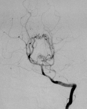
Cerebral vessel angiography shows arterial wall irregularities and focal stenotic lesions.
‘‘Moyamoya’’ comes from a Japanese term meaning ‘‘puff of smoke" and is an example of late onset RT induced vasculopathy.
Describes the angiographic appearance of collateralized vessels that form in the region of the basal ganglia and beyond when RT or other vasculopathy occludes the
internal carotid or proximal anterior and middle cerebral arteries
- Long term problem after RT especially in NF1 patients
- Progressive narrowing of the brain’s blood vessels, and obliteration of the vessels in the circle of Willis
Epidemiology:
- Often occurs in patients treated for optic nerve glioma
- Young children less than 5 years old with neurofibromatosis type 1 (NF-1) are most at risk
- RT dose of more than 5000 cGy increases the risk
- Syndrome usually develops within 8 years of therapy
- Adults with RT induced vasculopathy are more likely to present with intra-cranial hemorrhage.
Symptoms:
- Transient ischemic attacks
- Cerebrovascular events (strokes)
- Motor weakness
- Dementia
Pathology:
- Basilar occlusive disease occurs in:
- Large arteries of the circle of Willis
- Terminal parts of the internal carotid arteries
- Proximal portions of the middle cerebral artery
- Anterior cerebral artery
- Most frequent area for this to occur is near the sella
- Stenosis is often gradual so an extensive collateral circulation develops
Imaging:
- Angiographic appearance of telangiectatic vessels is like a “puff of smoke” or “misty” – which means moyamoya in Japanese.
Treatment:
- EDAS = EncephaloDuroArterioSynangiosis
- Transposition of a segment of the scalp artery on to the surface of the brain to improve collateral flow.
Study looking at the incidence of vascular disease in craniopharyngioma patients given RT (1995 to 2008) at a single center15.
- 22 patients
- 20 given RT had imaging available.
- 6 of the 20 had some type of vasculopathy
- 1 - bilateral temporal cavernomas
- 1 - moyamoya syndrome
- 1 - aneurysm of the internal carotid artery
- 3 - decreased caliber of the carotid/cerebral arteries (asymptomatic)
- 2 of 6 children with abnormalities also had intracystic bleomycin prior to RT