Dental
RT related Damage
The risk of radiation therapy (RT) induced damage depends on many factors, but the following are very important:
- Type of radiation:
- Orthovoltage RT was used to treat patients many years ago. The dose from orthovoltage is magnified in bone, resulting in increased damage to bone.
- Dose and fractionation of RT:
- The higher doses of RT are associated with an increased risk of damage
- Age of child at time of RT:
- The younger the child, then the more potential there is for long term damage and growth disturbance
Abnormal Dental Development:
Dental defects may include:
- Tooth and root under-development
- small teeth (dwarfism)
- root blunting/tapering
- Abnormal development of enamel and incomplete calcification
- teeth can be discoloured or have white patches
- grooves or pits in teeth
- increased risk of dental caries
- Premature apical closure of roots
- Agenesis of teeth
- Delayed eruption or impaction of teeth
- Malocclusion (bite problem with overbite or underbite)
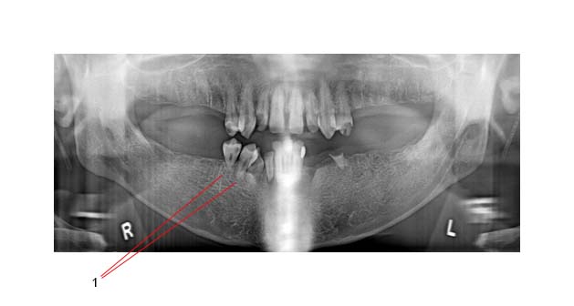
The panorex image below shows hypoplastic root development (1) after RT as a 7 year old child:

Odontogenic cell sensitivity:
- Depends on the cell cycle and the mitotic activity of odontoblasts at the time of the radiation exposure.
- Odontoblasts are most susceptible to RT damage when they are actively proliferating as they lay down the dentin matrix of teeth.
- RT at this critical time will stop the mitotic activity of odontoblasts and the result is dentin that is not effectively bonded to enamel.
Age and RT dose:
- The effect of RT is most pronounced when the child is less than five years old at the time of therapy. In a very young child, doses greater than 4 Gy may cause permanent localized dental defects.
- It was initially thought that it took 10 Gy to permanently damage ameloblasts (the cells responsible for enamel formation) while 30 Gy was needed to arrest tooth development1. However, if chemotherapy is combined with RT, a much lower RT dose is needed to have the same destructive effects1.
- Doses between 10-18 Gy of RT (when administered late in tooth formation) can cause a distortion in root apexification.
- Higher RT doses than 20 Gy during earlier stages of dental development may result in complete agenesis.
- Long-term studies have shown a slightly increased risk of dental problems after exposure to RT doses of up to 20 Gy (OR 1.3), but that the risk is much higher with doses greater than 20 Gy (OR 5.6). Likewise, the risk of oral soft tissue damage is low with RT doses less than 20 Gy (OR 1.2) but higher with doses greater than 20 Gy (OR 2.2)3.
- When developing teeth are in a high-dose RT field, the apices tend to close earlier than normal resulting in teeth with shorter roots. The morphology of the roots may also be affected.
Xerostomia
Salivary gland dysfunction can occur at much lower RT doses.
When salivary glands are permanently damaged, xerostomia can result which predisposes the patient to dental decay2,3,4.
Current research shows that only 25% of the pre-RT stimulated saliva was present when both parotid glands receive a mean dose of 32 Gy. The risk of xerostomia increases significantly if the RT dose to the parotid glands exceeds 40 Gy.
Salivary flow rate was also reduced by approximately 4% per Gy of the mean parotid dose8.
Patients who receive high dose RT to their salivary glands have markedly reduced salivary flow resulting in a increased risk of dental decay1.
“Radiation caries” is characterized by decay which starts at the neck (or cervical third) of the tooth. The decay then progresses inward into the dentin and cementum, ultimately undermining the strength of the tooth1.
Because of the xerostomia, there is an increased risk of periodontal (gum) disease. Gums can shrink away from the teeth causing infection around the roots of the teeth and this leads to bone loss and "loosening" of the teeth.
Trismus
Underlying disease and high dose RT to the face may lead to fibrosis (scarring) and damage to the temporo-mandibular joint (TMJ). This can cause trismus and an inability to open the mouth very wide. Therefore it can be very difficult to maintain oral hygiene in these circumstances and periodontal disease can become a serious problem.
Patients may also have temporo-mandibular joint dysfunction with pain in the TMJ.
Second Cancers
After RT there is always a small risk of the development of secondary oral cancers many years after treatment within the previous RT treatment field.
Survivors after BMT are especially at risk for this complication.
This is a rare condition (risk probably less than 3%) and can occur in patients who have dental surgery years after high dose RT to the mandible. It means that dead (or necrotic) bone develops after injury.
This would be rare in patients who received less than 60 Gy.
It is due most likely to damage to the small blood vessels by the RT. The bone is unable to repair damage due to trauma. The inferior alveolar artery (the predominant arterial blood supply to the body of the mandible) and periosteal arteries have significant intimal fibrosis and thrombosis
Presentation:
- Pain and swelling
- Exposed bone
- Pathologic fracture
- Oral cutaneous fistula formation
The severity of this problem is "graded"
- Grade I: exposed alveolar bone
- Grade II: exposed alveolar bone that does not respond to HBO
- Grade III: full-thickness involvement and/or pathologic fracture
Children's Oncology Group survivorship guidelines: Osteoradionecrosis

